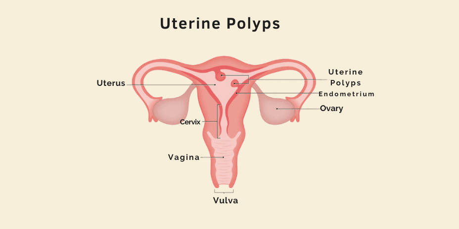
Uterine polyp is a common condition affecting over 1 million people every year in India. They are benign growths attached to the uterine lining. Overgrown endometrium cells lead to uterine polyps. They are also called endometrial polyps. It is possible to have more than one polyp. The polyps formed can range from a few millimeters to a few centimeters. They generally occur within the uterus, but sometimes they may slip into the vagina through the cervix—the lowermost part of the uterus.
CAUSES
The exact cause of uterine polyps is not known. But studies link them to the following:
Estrogen levels: A high level of estrogen can cause uterine polyps.
Inflammation: Prolong inflammation of female reproductive system parts like the uterus, vagina, and cervix.
RISK FACTORS
Risk factors mainly include the factors that increase estrogen levels.
Related to high estrogen levels:
- Pregnancies & Childbearing years: Estrogen levels are incredibly high during pregnancies and childbearing years.
- Pre-menopausal age: After reaching menopause, estrogen levels drop, but just before menopause, they spike.
- Obesity: Fat cells produce estrogen.
Others:
- High blood pressure.
- Miscarriage
- Herpes
- HPV infection
SYMPTOMS
Almost all symptoms of uterine polyps are related to the menstrual cycle.
- Bleeding between menstrual cycles.
- Bleeding after menopause
- Irregular menstrual periods
- Severe menstrual periods
- Infertility
DIAGNOSIS
The diagnosis may involve a pelvic exam, ultrasound, hysterosonogram, and endometrial biopsy.
Pelvic Examination: A pelvic examination is a routine screening test in which the doctor examines various organs, such as the vulva, ovaries, and uterus, from outside. The doctor may also use a speculum, a device that helps the doctor view the patient’s cervix and inspect it for abnormalities.
Transvaginal Ultrasound: Ultrasound is an imaging technique that uses ultrasound waves to create an image/video of the patient’s internal organs. In transvaginal ultrasound, the doctor will insert the transducer into the patient’s vagina to inspect the uterus with the help of the video/image on screen.
Hysterosonogram: In some cases, ultrasound may not be able to show the complications. Hence, to ascertain polyps, the doctor may recommend a hysterosonogram. This procedure is similar to ultrasound, but in this procedure, the doctor also inserts a saline solution into the cavity of the uterus. This helps the doctor see things that would not be possible with normal ultrasound.
Endometrial Biopsy: Sometimes, the doctor may recommend an endometrial biopsy. Here, the doctor will try to obtain sample tissues from the uterine lining for analysis. Most patients do not require anesthesia.
TREATMENT
Depending on the size of the polyp, the doctor may recommend one of the following treatments.
Watchful waiting: Polyps that are small in size may go away themselves. There is no reason to treat small polyps unless they are cancerous.
Medication: Medications can provide relief from symptoms, but such relief is likely to be temporary because, in most cases, the symptoms return when the medication is stopped.
Hysteroscopy: Hysteroscopy is a procedure in which the doctor inserts a small, lighted instrument known as the hysteroscope through the patient’s natural orifices. This instrument relays images on the screen, which helps the doctor inspect the patient’s inner organs and remove the polyps.


