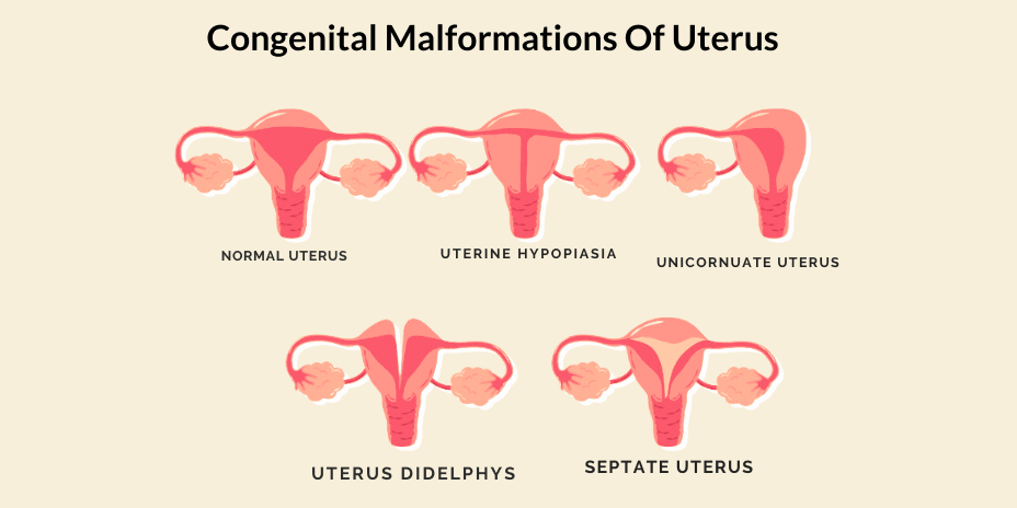
A condition is said to be a congenital anomaly when it affects a person from birth. It is estimated that 6.7% of the general population suffer from such malformations of the uterus, while the incidence of congenital malformation in women suffering from infertility is 7.4%. This incidence is significantly higher in women with a history of recurrent miscarriages.
Types of congenital malformation of the uterus:
Müllerian agenesis: Müllerian agenesis is a condition which occurs when the uterus, upper vagina and cervix (opening of the uterus) don’t develop correctly or at all. Women with this condition develop normal ovaries, breasts, clitoris and vulva.
SYMPTOMS
- Lack of menstrual cycle during puberty
- Pain during intercourse
- Renal anomalies
- Kidney problems
- Bone malformations
DIAGNOSES
The diagnoses may involve one of the following imaging techniques:
- MRI
- Ultrasound
- Sonohysterography
- X-ray
MANAGEMENT
It may be possible to conceive with a unicornuate uterus. Still, it needs to be managed carefully since cases of premature birth, pregnancy loss, malpresentation, and fetal demise are more common in such pregnancies. A c-section may also be required instead of a normal pregnancy.
Uterus didelphys: Uterine didelphys, also known as double uterus, is a rare congenital uterus anomaly. It occurs during the fetus’s development stage when the small tubes that normally join to form the uterus develop into two separate structures (two uteruses). One cervix (mouth of the uterus) is possible, or each structure may have its separate cervix.
SYMPTOMS
Women suffering from this condition usually do not feel any symptoms, but some women may experience:
- Menstrual bleeding does not stop even after using tampons
- Miscarriages
General & Pregnancy Complications
- Infertility
- Miscarriage
- Premature birth
- Kidney abnormalities
DIAGNOSIS
Diagnosis is carried out using various imaging techniques such as:
- Ultrasound
- Sonohysterogram
- Magnetic resonance imaging (MRI).
- Hysterosalpingography
Treatment :
Surgery can result in better management of pregnancies, other than that it is not medically necessary to undergo treatment unless the patient is experiencing severe symptoms.
BICORNUATE UTERUS
Bicornuate uterus, also known as heart-shaped uterus, is a condition in which the uterus has two horns separated by a septum. This condition is one of the most common uterus anomalies, along with septate uterus. It results from improper uterus formation during the prenatal years. This can happen for several reasons, one of them being exposure to diethylstilbestrol (DES).
Symptoms :
Women having a bicornuate uterus usually don’t experience any symptoms other than infertility.
Diagnosis :
To ascertain a bicornuate uterus, various imaging techniques or diagnostic laparoscopies:
- MRI
- 3D Ultrasound
- HSG
- Laparoscopy
Effect on pregnancy
A bicornuate uterus is known to have the following adverse effects on pregnancy:
- Preterm labour
- Cervical insufficiency
- Miscarriage in 2nd trimester
UTERINE Septum
In this condition, extra tissue called the septum hangs from the top of the uterus. The septum is often shaped like a wedge and may be small or hang from the top to the cervix (mouth of the uterus), thereby dividing the uterus into two cavities.
Causes :
Since the uterine septum is a congenital anomaly, women affected by it are born with it. It is developed due to the failure of the two uteri to absorb the dividing interior wall.
Symptoms :
The uterine septum shows no symptoms except infertility. Most women affected by uterine septum discover they have it when trying to understand the reason behind their infertility.
Diagnoses :
Various imaging techniques may be used to ascertain uterine septum:
- Transvaginal Ultrasound
- HSG:
- MRI:
Treatment :
- Hysteroscopic Resection: The septum is resected with the help of a hysteroscopy.


